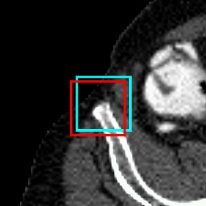
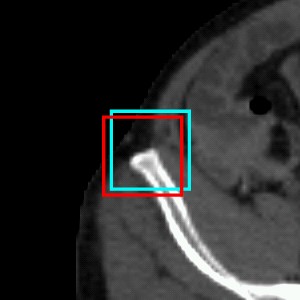
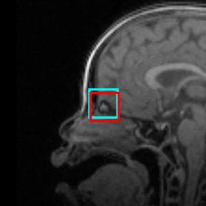
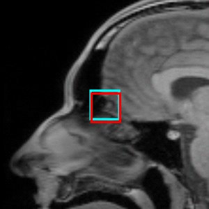
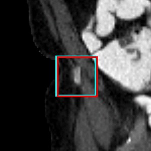
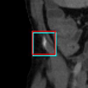
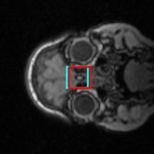
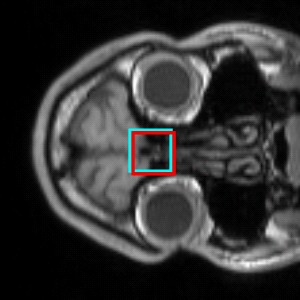
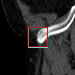
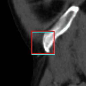
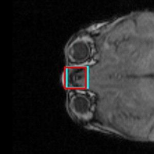
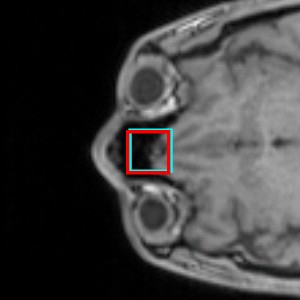
| Progressive Data Transmission for Hierarchical Detection in a Cloud (DiC) This page gives a high level overview of our research on Detection in a Cloud (DiC). For more details, please refer to our articles published in HP-MICCAI 2010 proceedings and Methods of Information in Medicine journal. Contents Overview In response to the growing need for image analysis services in the cloud computing environment, this paper proposes an automatic system for detecting landmarks in 3D volumes. The inherent problem of limited bandwidth between a (thin) client, Data Center (DC), and Data Analysis (DA) server is addressed by a hierarchical detection algorithm that obtains data by progressively transmitting only image regions required for processing. The client sends a request for a visualization of a specific landmark. The algorithm obtains a coarse level image from DC and outputs landmark location candidates. The coarse landmark location candidates are then used to obtain image neighborhood regions at a finer resolution level. The final location is computed as the robust mean of the strongest candidates after refinement at the subsequent resolution levels. The feedback about candidates detected at a coarser resolution makes it possible to only transmit image regions surrounding these candidates at a finer resolution rather then the entire images. Furthermore, the image regions are lossy compressed with JPEG 2000. Together, these properties amount to at least 50 times bandwidth reduction while achieving similar accuracy when compared to an algorithm using the original data. Motivation and Intuition Figure 1: The Detection in a Cloud (DiC) system is used by thin-client devices that request the display of an anatomical part for a specific patient. The patient data stored in a Data Center are transmitted to a high performance Data Analysis server that runs the detection algorithm. (In the medical domain and also in this paper, these servers are referred to as the Picture Archiving and Communication System (PACS) and Computer Aided Detection (CAD) server, respectively). The image with the anatomy highlighted is returned back to the client for display (Figure 1). 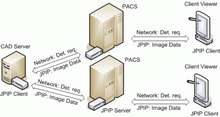 | We propose an efficient hierarchical learning-based detection system to avoid the problem of transmitting large datasets. The system runs on the CAD server that obtains portions of the original dataset from the PACS server on demand. The algorithm starts detection on a downsampled low-resolution image that has been compressed and transmitted to the CAD server. The coarse landmark candidate positions define the regions in a finer resolution image, where the coarse candidates are refined. The refinement steps continue until all levels of the hierarchy have been processed. The final detection result is obtained by robustly combining strongest candidates from the finest level. Figure 2: Overall DiC system diagram. The hierarchical detection algorithm progressively obtains image regions required for detection at each level. 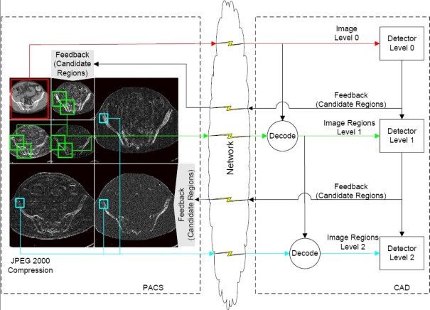 | The amount of transmitted data is significantly reduced in the DiC system. First, the algorithm only processes candidate regions at finer resolutions rather than the entire images. Second, all image regions are compressed with a lossy compression. When combined, these properties result in an overall reduction of the original data size by a factor of 30 (CT data) and by a factor of 196 (MRI data). Our experiments show that the lossy compression does not hinder the final detection accuracy. The experiments also demonstrate the robustness and accuracy of the hierarchical algorithm and advantages of training on compressed images. In summary, the paper makes three main contributions: (1) an overall system for landmark detection using remote datasets, (2) hierarchical detection algorithm with a local refinement, and (3) evaluation of training and detection on images compressed with lossy 3D JPEG 2000. Results Figure 3: Detection error vs. average volume size for hip bone landmark in CT (top) and crista galli landmark in brain MRI (bottom). The images were compressed in training and testing with the same pSNR level (adaptive) and uncompressed in training and compressed in testing (nonadaptive). The hierarchical processing results in lowest detection error through the focused coarse-to-fine search and training on compressed volumes. The average size of the uncompressed and lossless-compressed volumes is: 404 kB and 189 kB (8 mm CT), 3334 kB and 985 kB (2 mm MRI). 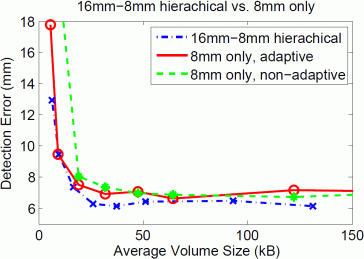 | 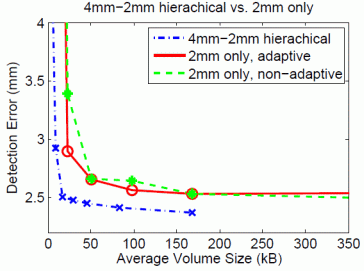 | Table 1: The median detection error of the hierarchical detection on images compressed at pSNR 70 (2nd and 5th column), on uncompressed images (3rd and 6th column), and on a single resolution losslessly-compressed images (4th and 7th column). The average size of uncompressed volumes is 3188 kB (4 mm CT) and 3334 kB (2 mm MRI). The hierarchical algorithm trained with images of pSNR 70 requires the least amount of data without sacrificing the detection accuracy. | | CT | MRI | | | 16-8-4 hier | 16-8-4 hier | 4 mm | 4-2 hier | 4-2 hier | 2 mm | | | pSNR 70 | lossless | lossless | pSNR 70 | lossless | lossless | | Error (mm) | 3.87 | 3.54 | 3.98 | 2.50 | 2.37 | 2.27 | | Avg. Data Size (kB) | 106.22 | 393.45 | 1345.96 | 16.99 | 168.07 | 984.76 | Summary At the core of the Detection in a Cloud (DiC) is a hierarchical learning algorithm that propagates position candidate hypotheses across a hierarchy of classifiers during training and detection. The algorithm only requires image regions surrounding the candidates which results in less bandwidth for remote data access. Further bandwidth savings (without sacrificing the detection accuracy) are achieved by compressing the images regions with lossy JPEG 2000. The total bandwidth savings for retrieving remotely stored data amount to 30 times (CT data) and 196.2 times (MRI data) reduction when compared to the original data size and 12.7 times (CT) and 58.0 (MRI) when compared to data size after lossless compression. The proposed approach makes it possible to shift the integration, maintenance, and software updates from the client to the CAD server. Therefore, when the classifiers are updated, they are immediately available to all clients. In the clinical environment, detected anatomical parts can be reviewed on the client devices remotely. The current system opens many exciting future research directions both on the algorithmic side as well as on the systems side. We are interested the most in building more complicated models with several landmarks of interest trained for different modalities. Such large scale systems will require coordination of multiple CAD servers possibly distributed in a wide-area network.
Publications and Further Reading -
  Sofka, M., Ralovich, K., Zhang, J., Zhou, S.K., Comaniciu, D., 2012. Progressive Data Transmission for Anatomical Landmark Detection in a Cloud. Methods of Information in Medicine 51, 268–278. Sofka, M., Ralovich, K., Zhang, J., Zhou, S.K., Comaniciu, D., 2012. Progressive Data Transmission for Anatomical Landmark Detection in a Cloud. Methods of Information in Medicine 51, 268–278. Background: In the concept of cloud-computing-based systems, various authorized users have secure access to patient records from a number of care delivery organizations from any location. This creates a growing need for remote visualization, advanced image processing, state-of-the-art image analysis, and computer aided diagnosis. Objectives: This paper proposes a system of algorithms for automatic detection of anatomical landmarks in 3D volumes in the cloud computing environment. The system addresses the inherent problem of limited bandwidth between a (thin) client, data center, and data analysis server. Methods: The problem of limited bandwidth is solved by a hierarchical sequential detection algorithm that obtains data by progressively transmitting only image regions required for processing. The client sends a request to detect a set of landmarks for region visualization or further analysis. The algorithm running on the data analysis server obtains a coarse level image from the data center and generates landmark location candidates. The candidates are then used to obtain image neighborhood regions at a finer resolution level for further detection. This way, the landmark locations are hierarchically and sequentially detected and refined. Results: Only image regions surrounding landmark location candidates need to be transmitted during detection. Furthermore, the image regions are lossy compressed with JPEG 2000. Together, these properties amount to at least 30 times bandwidth reduction while achieving similar accuracy when compared to an algorithm using the original data. Conclusions: The hierarchical sequential algorithm with progressive data transmission considerably reduces bandwidth requirements in cloud-based detection systems. @article{sofka:mim12,
author = {Sofka, Michal and Ralovich, Kristof and Zhang, Jingdan and Zhou, S.~Kevin and Comaniciu, Dorin},
title = {Progressive Data Transmission for Anatomical Landmark Detection in a Cloud},
journal = {Methods of Information in Medicine},
year = {2012},
note = {Invited Paper.},
volume = {51},
number = {3},
pages = {268--278},
keywords = {Cloud Computing, Machine Learning, Pattern Recognition System, Computer-Assisted Image Processing, Image Compression}
}
-
  Sofka, M., Ralovich, K., Zhang, J., Zhou, S.K., Comaniciu, D., 2010. Progressive Data Transmission for Hierarchical Detection in a Cloud. In: Proceedings of the 2nd International Workshop on High-Performance Medical Image Computing for Image-Assisted Clinical Intervention and Decision-Making (HP-MICCAI 2010). Bejing, China. Sofka, M., Ralovich, K., Zhang, J., Zhou, S.K., Comaniciu, D., 2010. Progressive Data Transmission for Hierarchical Detection in a Cloud. In: Proceedings of the 2nd International Workshop on High-Performance Medical Image Computing for Image-Assisted Clinical Intervention and Decision-Making (HP-MICCAI 2010). Bejing, China. In response to the growing need for image analysis services in the cloud computing environment, this paper proposes an automatic system for detecting landmarks in 3D volumes. The inherent problem of limited bandwidth between a (thin) client, Data Center (DC), and Data Analysis (DA) server is addressed by a hierarchical detection algorithm that obtains data by progressively transmitting only image regions required for processing. The client sends a request for a visualization of a specific landmark. The algorithm obtains a coarse level image from DC and outputs landmark location candidates. The coarse landmark location candidates are then used to obtain image neighborhood regions at a finer resolution level. The final location is computed as the robust mean of the strongest candidates after refinement at the subsequent resolution levels. The feedback about candidates detected at a coarser resolution makes it possible to only transmit image regions surrounding these candidates at a finer resolution rather then the entire images. Furthermore, the image regions are lossy compressed with JPEG 2000. Together, these properties amount to at least 50 times bandwidth reduction while achieving similar accuracy when compared to an algorithm using the original data. @inproceedings{sofka:HPmiccai10,
author = {Sofka, Michal and Ralovich, Kristof and Zhang, Jingdan and Zhou, S.~Kevin and Comaniciu, Dorin},
title = {Progressive Data Transmission for Hierarchical Detection in a Cloud},
booktitle = {Proceedings of the 2nd International Workshop on High-Performance
Medical Image Computing for Image-Assisted Clinical Intervention
and Decision-Making (HP-MICCAI 2010)},
year = {2010},
month = "24~" # sep,
address = {Bejing, China},
pages = {},
note = {\textbf{Best paper award.}}
}
|


